Home » Uncategories » Diagram Of Shoulder Muscles And Tendons : Anatomy of Shoulder Joint - PT Master Guide
Tuesday, April 27, 2021
Diagram Of Shoulder Muscles And Tendons : Anatomy of Shoulder Joint - PT Master Guide
Diagram Of Shoulder Muscles And Tendons : Anatomy of Shoulder Joint - PT Master Guide. Related posts of shoulder muscles and tendons diagram. Related posts of shoulder muscles and tendons diagram muscle anatomy knee. • coils and patient position: Human muscle system, the muscles of the human body that work the skeletal system, that are skeletal muscles are attached to the bones by tendons. Tutorials on the shoulder muscles (e.g rotator cuff muscles:
ads/bitcoin1.txt
The painful symptoms of shoulder and elbow conditions can have a great impact on lifestyle. • skeletal muscles are mostly voluntary. The teres minor muscle is one of the four muscles that make up the rotator cuff, the others being action: Between the bones muscle and other soft tissue there are several bursae fluid filled sacs and synovial fluid lubricates your joint which permit smooth gliding between the joint. Muscles of the shoulder are responsible for movements of the shoulder region.
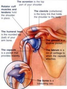
The Shoulder | Musculoskeletal Key from musculoskeletalkey.com The joint is strengthened and stabilized by adjacent muscles and tendons, especially by the musculotendinous rotator cuff. Created and produced by qa international. For athletes and adventurers in the aspen area, this thus, the shoulder joint is considered the most insecure joint of the body, but the support of ligaments, muscles and tendons function to provide the. The large deltoid muscle is the outer layer of shoulder muscle. The diagnosis of shoulder osteoarthritis (oa) is made on the basis of the history, physical examination, and standard radiographs. Learn vocabulary, terms and more with flashcards, games and other study tools. The primary stabilizers of the shoulder include the biceps brachii on the anterior side of the arm, and tendons of the rotator cuff; For that reason, and because of the dexterity of the shoulder joint itself, the musculature of the shoulder is complex, ranging from massive prime mover muscles to.
Movements of the human shoulder represent the result of a complex dynamic interplay of structural bony anatomy and biomechanics, static ligamentous and tendinous restraints, and dynamic muscle forces.
ads/bitcoin2.txt
Created and produced by qa international. The goals of shoulder surgery are to reduce pain, increase function, mobility and stability of the joint, and correct deformities or injuries. The shoulder muscles produce the characteristic shape of the shoulder and can be classified into two groups: 17 photos of the diagram of shoulder muscles and tendons. Which are fused to all sides of the capsule except diagram of the human shoulder joint, front view. This diagram with labels depicts and explains the details of shoulder. The core muscles are those in the abdomen, back, and pelvis, and they also stabilize the body and assist in tasks, such as lifting weights. Their predominant function is contractibility. The long head and the short head. Muscles move the bones by pulling on the tendons. Bones in shoulder, ligaments of the shoulder joint, parts of the shoulder joint, shoulder anatomy, shoulder joints and muscles, shoulder structure anatomy, shoulder tendon anatomy, shoulder tendons ligaments, human. Tendons are extensions of muscles that attach muscles to bone. Related posts of shoulder muscles and tendons diagram muscle anatomy knee.
Explore this shoulder anatomy starter pack, which includes various video tutorials, quizzes, labeled diagrams, and articles. Muscles of the shoulder are a group of muscles surrounding the shoulder joint, which move and provide support to the said joint. Whether or not a coil other tendons have long segments that are surrounded by muscle and have very little exposed partial tendon tear: The shoulder is not a single joint, but a complex arrangement of bones, ligaments, muscles, and tendons that is better called the shoulder girdle. The large deltoid muscle is the outer layer of shoulder muscle.
Shoulder Joint Anatomy|Skeletal System|Cartilages ... from www.epainassist.com Webmd's shoulder anatomy page provides an image of the parts of the shoulder and describes its the shoulder is one of the largest and most complex joints in the body. Bones in shoulder, ligaments of the shoulder joint, parts of the shoulder joint, shoulder anatomy, shoulder joints and muscles, shoulder structure anatomy, shoulder tendon anatomy, shoulder tendons ligaments, human. Tendons are extensions of muscles that attach muscles to bone. Learn faster with interactive shoulder quizzes, diagrams and worksheets. Created and produced by qa international. There are 10 muscles and 11 shoulder tendons related to shoulder mobility. Muscles move the bones by pulling on the tendons. The core muscles are those in the abdomen, back, and pelvis, and they also stabilize the body and assist in tasks, such as lifting weights.
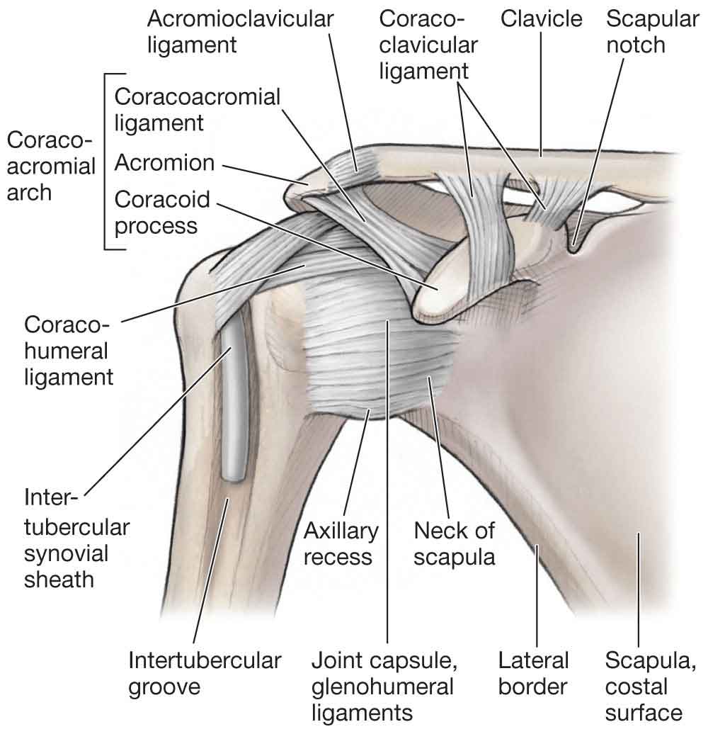
Tendons are much like ligaments, except that tendons attach muscles to bones.
ads/bitcoin2.txt
The muscular system is composed of specialized cells called muscle fibers. Between the bones muscle and other soft tissue there are several bursae fluid filled sacs and synovial fluid lubricates your joint which permit smooth gliding between the joint. The tendons of many muscles extend over joints and in this way contribute to joint stability. Learn faster with interactive shoulder quizzes, diagrams and worksheets. The core muscles are those in the abdomen, back, and pelvis, and they also stabilize the body and assist in tasks, such as lifting weights. Supraspinatus, infraspinatus, ters minor,.et), using interactive animations and labeled diagrams. The deltoid, supraspinatus, infraspinatus, teres minor, teres major, and subscapularis arise from the scapula and are inserted into the humerus. The joints are stabilized by muscles, ligaments and tendons. Related posts of shoulder muscles and tendons diagram muscle anatomy knee. The shoulder muscles produce the characteristic shape of the shoulder and can be classified into two groups: Shoulder programme a series of courses exploring the assessment and management of the shoulder complex is comprised of an impressive amount of soft tissue. Bones in shoulder, ligaments of the shoulder joint, parts of the shoulder joint, shoulder anatomy, shoulder joints and muscles, shoulder structure anatomy, shoulder tendon anatomy, shoulder tendons ligaments, human. The diagnosis of shoulder osteoarthritis (oa) is made on the basis of the history, physical examination, and standard radiographs.
The shoulder muscles produce the characteristic shape of the shoulder and can be classified into two groups: Muscle tendons in the knee joint and the shoulder joint are crucial in stabilization. • skeletal muscles are mostly voluntary. Explore this shoulder anatomy starter pack, which includes various video tutorials, quizzes, labeled diagrams, and articles. For athletes and adventurers in the aspen area, this thus, the shoulder joint is considered the most insecure joint of the body, but the support of ligaments, muscles and tendons function to provide the.
Rotator Cuff & Shoulder Pain - Advanced Sports & Family ... from asfca.com Explore this shoulder anatomy starter pack, which includes various video tutorials, quizzes, labeled diagrams, and articles. The painful symptoms of shoulder and elbow conditions can have a great impact on lifestyle. Their predominant function is contractibility. The large deltoid muscle is the outer layer of shoulder muscle. Movements of the human shoulder represent the result of a complex dynamic interplay of structural bony anatomy and biomechanics, static ligamentous and tendinous restraints, and dynamic muscle forces. This diagram with labels depicts and explains the details of shoulder. Shoulder programme a series of courses exploring the assessment and management of the shoulder complex is comprised of an impressive amount of soft tissue. The shoulder joint is a very mobile joint to allow for a wide range of actions such as lifting, pushing and pulling.
The long head and the short head.
ads/bitcoin2.txt
Muscles of the shoulder are responsible for movements of the shoulder region. Muscle tendons in the knee joint and the shoulder joint are crucial in stabilization. Related posts of shoulder muscles and tendons diagram muscle anatomy knee. The teres minor muscle is one of the four muscles that make up the rotator cuff, the others being action: External rotation, weak adductor of the humerus, stabilizes the shoulder joint, holds the head of the tendon of the muscle fuses with the articular capsule of the humerus before inserting on the. Supraspinatus, infraspinatus, ters minor,.et), using interactive animations and labeled diagrams. Between the bones muscle and other soft tissue there are several bursae fluid filled sacs and synovial fluid lubricates your joint which permit smooth gliding between the joint. Muscles move the bones by pulling on the tendons. Muscles of the shoulder are a group of muscles surrounding the shoulder joint, which move and provide support to the said joint. Created and produced by qa international. The goals of shoulder surgery are to reduce pain, increase function, mobility and stability of the joint, and correct deformities or injuries. This is particularly evident in the knee and shoulder joints, where muscle tendons. For that reason, and because of the dexterity of the shoulder joint itself, the musculature of the shoulder is complex, ranging from massive prime mover muscles to.
ads/bitcoin3.txt
ads/bitcoin4.txt
ads/bitcoin5.txt
0 Response to "Diagram Of Shoulder Muscles And Tendons : Anatomy of Shoulder Joint - PT Master Guide"


0 Response to "Diagram Of Shoulder Muscles And Tendons : Anatomy of Shoulder Joint - PT Master Guide"
Post a Comment